



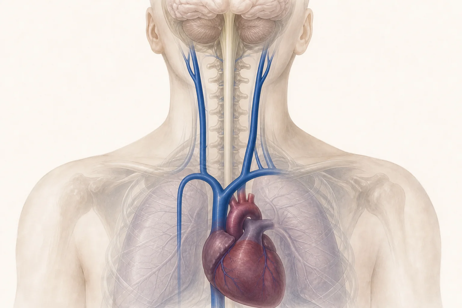
CCSVI, or Chronic Cerebrospinal Venous Insufficiency, is a clinical-haemodynamic hypothesis concerning venous drainage from the brain and spinal cord. This page reconstructs Dr Ciro Gargano’s experience in vascular diagnostics and angioplasty of the jugular and azygos veins, while illustrating what research has clarified in subsequent years.
An important clarification
CCSVI has been the subject of extensive scientific debate, particularly regarding its possible association with multiple sclerosis. Clinical experiences and individual patient testimonials are part of the history of this research, but they alone do not prove the effectiveness of a treatment. Venous angioplasty is not a cure for multiple sclerosis and does not replace neurological assessment or prescribed therapies.
On this page
What is CCSVI
The acronym CCSVI stands for Chronic Cerebrospinal Venous Insufficiency. Professor Paolo Zamboni proposed this term to describe a condition in which certain anomalies of the main extracranial venous pathways could hinder blood return from the central nervous system to the heart.
The most studied veins are the internal jugular veins, which drain a significant part of the blood from the brain, and the azygos vein, located in the chest and also connected to the venous drainage of the spinal cord. The described anomalies include dysplastic or poorly mobile valves, endoluminal membranes and septa, circular stenoses, hypoplasia of venous segments, and other anatomical or functional alterations.
The most scientifically debated point is not only the existence of individual venous anomalies, which can be documented, but their clinical significance: how much they affect drainage, how frequent they are in healthy people or those with other diseases, and what relationship they have, if any, with neurological symptoms.
Jugulars, azygos vein and possible outflow anomalies
In the venous system, flow depends on body position, respiration, pressure variations in the chest, and the availability of alternative venous pathways. For this reason, the evaluation of cerebrospinal drainage is more complex than simply measuring the diameter of a vein.
- Valvular anomalies: thickened, rigid, maloriented, or reduced mobility leaflets.
- Septa and membranes: endoluminal structures that can interfere with blood flow.
- Stenosis or annulus: focal or circular narrowing of the venous lumen.
- Hypoplasia: reduced development of a vein segment.
- Torsions or compressions: alterations in course or calibre that can vary with posture.
- Extrinsic compressions: sometimes structures external to the vein can compress it. Described muscular causes include anatomical variants or hypertrophy of the omohyoid muscle, which can cause compression of the internal jugular vein (PubMed insight).
- Agenesis: failure of development of a venous segment, with possible compensation through alternative venous pathways.
The mere presence of one of these characteristics does not automatically allow a neurological disorder to be attributed to it. Anatomy, haemodynamics, symptoms, and differential diagnosis must be considered separately.
The 2009 IUP Consensus and truncal venous malformations
In September 2009, an international panel of the IUP, International Union of Phlebology, unanimously recommended the use of the Hamburg classification for venous malformations. In the document, stenotic lesions of the main extracranial venous pathways — including jugulars, superior caval system, and azygos vein — were classified as congenital truncal venous malformations.
It is therefore correct to remember that the consensus document recognised and classified this type of venous anomaly. It is more precise, however, not to transform this passage into a definitive approval of the entire CCSVI theory or its treatment in multiple sclerosis: these aspects have been evaluated by subsequent studies and remain conceptually distinct.
Read the IUP consensus document indexed in PubMed.
The colour duplex ultrasound protocol proposed by Professor Zamboni
The original protocol involved a venous colour duplex ultrasound performed with a specific methodology, in supine and seated positions, to search for five haemodynamic criteria:
- reflux in the internal jugular and/or vertebral veins in different positions;
- reflux in the deep cerebral veins;
- ultrasound evidence of anomalies or narrowing of the internal jugulars;
- absence of detectable flow in the internal jugular and/or vertebral veins;
- alteration of normal postural control of cerebral venous outflow.
According to Zamboni’s proposal, the presence of at least two out of five criteria allowed the examination to be classified as positive for CCSVI. Dr Gargano trained on this protocol and applied it in the historical diagnostic pathway of his patients.
Subsequent studies revealed reproducibility issues between operators and centres. Colour duplex ultrasound is, in fact, a dynamic and operator-dependent examination; technique, position, respiration, and interpretive criteria can alter the result. The five criteria should therefore be presented as part of the historical research protocol, not as a universally accepted diagnostic standard today.
Further reading on the inter-operator reproducibility of the Doppler protocol.
What venous angioplasty consists of
Venous angioplasty for CCSVI is an endovascular procedure. Through a venous access, generally femoral, a catheter is introduced, and a phlebography is performed to study the affected veins. If a narrowing is identified, a balloon is positioned in the segment to be treated and inflated with the aim of enlarging the lumen.
In the period documented by the old website, Dr Gargano’s protocol included colour duplex ultrasound control before, during, and after the procedure, and a scheduled clinical re-evaluation. The historical choice was to use the balloon without implanting stents in the jugular veins, in the absence of dedicated venous devices and considering the possible risks of implantation in that site.
Like all invasive procedures, venoplasty can also involve complications. Indication, expected benefits, alternatives, and risks must be evaluated based on available evidence and current clinical recommendations.
Dr Ciro Gargano’s training and experience
CCSVI represented an important part of Dr Ciro Gargano’s professional and scientific activity. In the academic year 2010/2011, he obtained a Second Level University Master’s Degree in Haemodynamics and Advanced Venous Surgery from the University of Ferrara, lasting 60 ECTS credits, passing the final examination on 20 October 2011.
The successful completion of the examination was certified by the Vascular Diseases Centre of the University of Ferrara with a declaration signed by Professor Paolo Zamboni as the Centre’s director. Following this training, Dr Gargano managed a personal caseload of over 1,500 procedures on the jugular and azygos veins.
The activity was not limited to the technical execution of angioplasty. The pathway was organised in collaboration with other professionals and included vascular, neurological, and neuropsychological evaluation, ultrasound controls, and follow-up. This approach aimed to observe any clinical variations in a structured manner, without reducing them to the patient’s immediate impression alone.
The multidisciplinary protocol and neuropsychological evaluation
The historical protocol documented by the Centre included pre-intervention evaluations, controls during and immediately after the procedure, and a long-term re-evaluation. The neuropsychological battery used included, according to clinical needs:
- Selective Reminding Test;
- 10/36 Spatial Recall Test;
- Symbol Digit Modalities Test;
- Paced Auditory Serial Addition Test (PASAT);
- delayed recall tests;
- Word List Generation.
Questionnaires relating to mood, quality of life, and disease impact were also used, including BDI-II, PHQ-9, MSQOL-54, and MSIS-29; for caregivers, CAREQOL-MS was also provided. The value of this documentation lies primarily in having adopted repeatable measures for cognitive functions and quality of life.
The observational study: 53 patients and six-month follow-up
In March 2011, Dr Gargano initiated the “Observational, non-profit study, aimed at evaluating the EDSS (Expanded Disability Status Score) of patients with multiple sclerosis and chronic cerebrospinal insufficiency (CCSVI) treated with percutaneous angiodilatation”. The accompanying documentation preserved reports opinion no. 2/r.p.s.o of 16 March 2011 and authorisation decree no. 8 of 21 March 2011 for the project. The protocol initially envisaged 40 subjects; between April and June 2011, 53 patients meeting the specified criteria were actually enrolled.
Evaluations were carried out before the intervention, after 30 days, and after six months. The primary outcome was EDSS; depression, impact of multiple sclerosis, and quality of life were also measured using BDI, MSIS-29, EQ-5D, and EQ-5D VAS. The mean age of the sample was 42.4 years, the mean disease duration 11.9 years; 37.7% had a relapsing-remitting form, 54.7% a secondary progressive form, and 7.6% a primary progressive form.
Observed EDSS values
| Evaluation | Mean EDSS | Patients evaluated | Comparison with initial value |
|---|---|---|---|
| Before intervention | 5.4 ± 2.1 | 53 | Baseline value |
| After 30 days | 4.8 ± 2.3 | 53 | p < 0.001 |
| After 180 days | 4.5 ± 2.4 | 44 | p < 0.001 |
In the conclusive report, patients with EDSS reduction were classified as “responders”, while those with stable or increased EDSS were classified as “non-responders”. According to this definition, the report indicated a responder rate of 64.2% (95% confidence interval: 50.7%-75.7%).
BDI, MSIS-29, EQ-5D, and EQ-5D VAS showed a mean improvement at 30 days in both groups. At six months, the improvement remained statistically significant only in the group classified as responders. The same report interpreted the initial improvement common to both groups as compatible with a possible short-term placebo effect.
Adverse events reported
The document recorded two venous wall haematomas, amounting to 3.8%, resolved in approximately two weeks; one venous thrombosis, amounting to 1.9%; and one venous restenosis, amounting to 1.9%. The authors specified that the sample size was not sufficient to reliably estimate the incidence of relatively rare events.
How the results should be interpreted
The report explicitly concluded that, due to the observational nature of the study, it was not possible to establish a cause-and-effect relationship between angioplasty and improvement in clinical conditions related to multiple sclerosis. Randomisation and a control group were lacking, and the number of participants was limited, especially for subgroup analysis and safety.
The value of the work therefore lies in having prospectively documented a real case series with standardised scales, pre-established control times, and recording of adverse events. The results represented an observational signal to be further investigated and must now be read together with subsequently published controlled studies.
Improvements reported by patients and their significance
Over the years, numerous patients have reported changes in fatigue, walking, sensation, sleep quality, thermoregulation, or bladder function. Some of these experiences are preserved in an extensive video archive created after the procedures.
These testimonials are important as a record of individual experiences and as a source of research questions. However, they do not represent proof of efficacy: they lack a control group, can be influenced by case selection, and do not allow prediction of the outcome for another person. The duration of a reported improvement can also vary considerably.
Consult the historical archive on the YouTube channel.
CCSVI and multiple sclerosis: what studies show today
To correctly interpret the literature, it is useful to distinguish three questions: whether certain venous anomalies are more frequent in people with multiple sclerosis; whether they play a causal role in the disease; whether correcting them with angioplasty produces a reliable clinical benefit.
Association
A meta-analysis published in 2023 in BMJ Open, comprising 20 studies and 3,069 participants, found a higher frequency of CCSVI in multiple sclerosis groups compared to healthy controls. The overall estimate was OR 3.36, but with high heterogeneity between studies (I² 79%). The result suggests a statistical association; it does not demonstrate that CCSVI is the cause of multiple sclerosis.
Read the meta-analysis published in BMJ Open.
Efficacy of angioplasty
The BRAVE DREAMS randomised controlled study did not demonstrate a general clinical benefit of venoplasty on the functional outcomes considered and concluded that the procedure could not be recommended for the treatment of multiple sclerosis. A subsequent broader analysis discussed possible radiological signals and differences related to venous morphology, but did not alter the general clinical recommendations.
The available Cochrane review found no benefits on primary patient-centred outcomes, including disability, relapses, quality of life, and physical or cognitive functions. The currently published NICE guidance states that venoplasty for CCSVI should not be used in the management of multiple sclerosis, given the absence of demonstrated benefit and possible complications.
- Cochrane review on venoplasty for CCSVI in multiple sclerosis
- Currently published NICE recommendations
The relationship with neurological therapies
Today, there is no therapy that guarantees a definitive cure for multiple sclerosis. However, there are disease-modifying treatments capable of reducing inflammatory activity, relapses, and, in many cases, the risk of progression. The choice depends on the type and activity of the disease and must be shared with the neurologist.
Vascular evaluation should not lead to independently stopping or modifying prescribed medications. This principle was already present in the FAQs of Dr Gargano’s old website and remains essential.
AISM information on therapies and treatments for multiple sclerosis.
A professional experience that deserves to be documented
The debate on CCSVI has been intense and often polarised. Documenting this experience today does not mean ignoring subsequent studies or erasing what has been observed in clinical practice. It means preserving a relevant part of Dr Gargano’s professional history, making protocols and materials available for consultation, and helping the reader distinguish between hypotheses, observations, testimonials, controlled studies, and recommendations.
The historical archive includes television and journalistic interviews, scientific documents, clinical cases, and numerous video testimonials. The content will be progressively selected and republished with the necessary context, avoiding technically obsolete materials or those lacking adequate documentation.
Frequently Asked Questions about CCSVI
What is CCSVI?
It is the name proposed for a condition characterised by anatomical or haemodynamic anomalies of the main venous pathways draining the brain and spinal cord. Its clinical significance and relationship with neurological diseases have been the subject of research and debate.
Which veins are involved?
The most studied are the internal jugular veins and the azygos vein. Vertebral veins and alternative venous pathways that participate in cerebrospinal drainage can also be evaluated.
How was it diagnosed according to the Zamboni protocol?
With a specific colour duplex ultrasound performed in supine and seated positions. The protocol sought five haemodynamic criteria and defined the examination as positive when at least two were present. However, the reproducibility of the method between operators and centres proved variable.
What did the 2009 IUP Consensus establish?
The panel unanimously recommended the Hamburg classification and included stenotic lesions of the extracranial venous pathways among congenital truncal venous malformations. This anatomical classification does not equate to a demonstration that CCSVI causes multiple sclerosis or that angioplasty cures it.
What does angioplasty of the jugular and azygos veins involve?
It is an endovascular procedure in which a catheter reaches the vein through peripheral access. After phlebographic study, a balloon can be inflated at the narrowed point to enlarge the lumen. It is an invasive procedure and not without risks.
Does CCSVI cause multiple sclerosis?
It has not been demonstrated. Some studies and a meta-analysis have found a statistical association, but with very heterogeneous results. An association does not establish which phenomenon comes first, nor does it prove a cause-and-effect relationship.
Does angioplasty cure multiple sclerosis?
No. Controlled studies have not demonstrated a general clinical benefit sufficient to recommend venoplasty in the management of multiple sclerosis. The procedure should not be presented as a cure or replace neurological therapies.
Why have some patients reported improvements?
Individual experiences can be genuine, but alone they do not allow the cause of the change to be identified. Fluctuating disease course, concomitant therapies, expectations, rehabilitation, case selection, and other factors can contribute. Controlled comparisons and standardised measures are necessary to attribute an effect to the procedure.
What was Dr Ciro Gargano’s experience?
Dr Gargano obtained a Second Level University Master’s Degree in Haemodynamics and Advanced Venous Surgery from the University of Ferrara, passing the final examination on 20 October 2011. The successful completion of the examination is attested by a declaration from the Vascular Diseases Centre signed by Professor Paolo Zamboni as director. He managed a personal caseload of over 1,500 procedures on the jugular and azygos veins, as part of a pathway that included vascular, neurological, and neuropsychological evaluation, ultrasound controls, and follow-up.
Where can historical testimonials and interviews be found?
Some of the videos are still available on the historical YouTube channel. Television interviews, articles, and documents from the old website are being catalogued and will be republished in a contextualised selection.
Last medical review: August 2026. Informational and historical content; does not replace a specialist visit or personalised neurological advice.
